Lump on Dog? Skin Tag, Cyst & Bump Checker — AI Photo Analysis
Found a lump on your dog? Upload a photo and get an instant AI health report. Identify skin tags, warts, cysts, lipomas, and other bumps to know if you need to see a vet.
Drop your pet's photo here
or
✅JPG, PNG, WEBP
📏Max 8MB
This tool provides AI-generated preliminary analysis only. Not a substitute for professional veterinary diagnosis.
📸 Photo Guide
Good photos
Close-up, clear
Shows shape & color
Avoid
Too far away
Blurry
Tips for best results
- ✓Part the fur to expose the lump fully before photographing
- ✓Take the photo in good lighting — natural daylight is best
- ✓Get close enough to see the texture, color, and borders of the lump
- ✓Include a coin or finger next to the lump for size reference
- ✓If the lump has multiple angles, take photos from different sides
How It Works — AI Dog Lump & Bump Checker

Step 1
Upload a Photo
Take a clear, close-up photo of the lump or bump on your dog. Include the surrounding skin for size comparison.

Step 2
AI Analyzes
Our AI examines the lump's shape, color, texture, border, and surrounding skin to identify potential conditions.

Step 3
Get Your Report
Receive a detailed health report with the likely condition, whether it may be benign or needs vet attention, and recommended next steps.
Common Lumps & Bumps on Dogs
Found a lump on your dog? Don't panic — most lumps on dogs are benign. But some need veterinary attention. Here are the most common types our AI can help identify. Also try our cat lump checker or dog skin checker or dog dental checker.
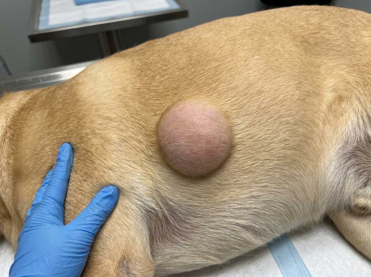
Lipoma (Fatty Lump)
Lipomas are the most common lumps found on dogs — soft, round, fatty tumors that sit just under the skin. They feel squishy and move freely when you push them. Lipomas in dogs are almost always benign and grow slowly over months or years. They're most common in middle-aged to older dogs, overweight dogs, and breeds like Labrador Retrievers, Dobermans, and Weimaraners. A soft lump on dog that moves easily is often a lipoma. They typically don't need removal unless they're in an awkward location (armpit, leg) that affects movement. How to shrink a dog lipoma naturally? Unfortunately, lipomas don't shrink on their own, but weight management can slow their growth. Your vet can confirm with a quick needle aspirate.
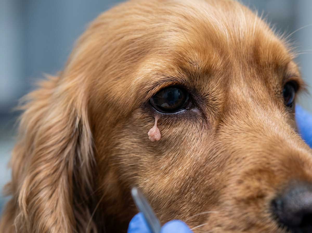
Skin Tag (Acrochordon)
Dog skin tags are small, soft, flesh-colored or slightly darker growths that hang from the skin on a thin stalk. They're extremely common in older dogs and completely harmless. Dog skin tag vs tumor? Skin tags are pedunculated (hang from a narrow base), soft, and don't grow over time — tumors are typically broader-based and may change. A common confusion is tick or skin tag on dog — check for tiny legs near the base to rule out a tick. Dog skin tag on eyelid is common and usually harmless unless it irritates the eye. Skin tags don't need removal unless they're snagged by collars or cause irritation. If a skin tag starts growing, bleeding without trauma, or changing color, have your vet check it.
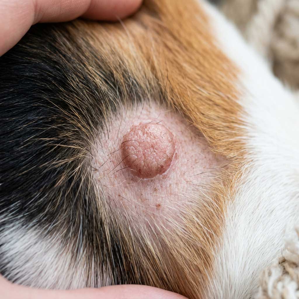
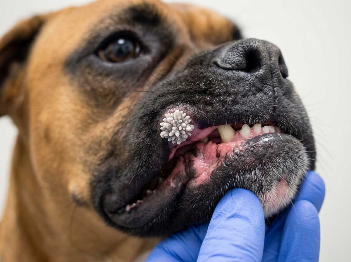
Warts (Papilloma)
Dog warts (papillomas) are caused by the canine papillomavirus. They look like small, rough, cauliflower-textured bumps — typically gray, white, or flesh-colored. Warts on dogs are most common in young dogs (under 2 years, whose immune systems are still developing) and immunocompromised older dogs. Dog mouth warts (oral papillomas) are especially common in puppies and usually resolve on their own within 1-5 months as the immune system fights off the virus. Warts from dogs are contagious to other dogs but NOT to humans. Cancerous wart on dog? True warts are benign, but squamous cell carcinomas can sometimes look wart-like — if a "wart" grows rapidly, ulcerates, or appears in an older dog, have it checked.
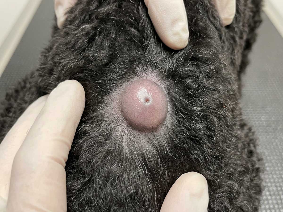
Sebaceous Cyst
A sebaceous cyst dog develops when a sebaceous (oil) gland becomes blocked, forming a round, raised, smooth lump filled with thick, white or yellowish material. They feel firm but slightly compressible — like a marble under the skin. Sebaceous cysts in dogs are benign and very common. Dog cyst burst is messy but not dangerous — you'll see thick, cheesy, foul-smelling discharge. Don't squeeze cysts yourself as this can cause infection. Dog cyst vs tumor? Cysts are typically round, well-defined, and may have a visible pore; tumors tend to be more irregular. Ruptured cyst on dog should be kept clean and monitored. Your vet may recommend removal if a cyst keeps rupturing or gets infected.
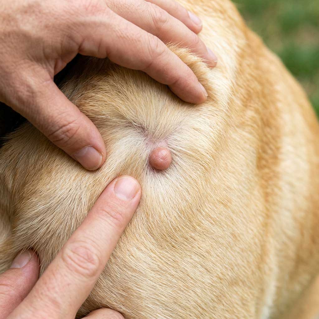
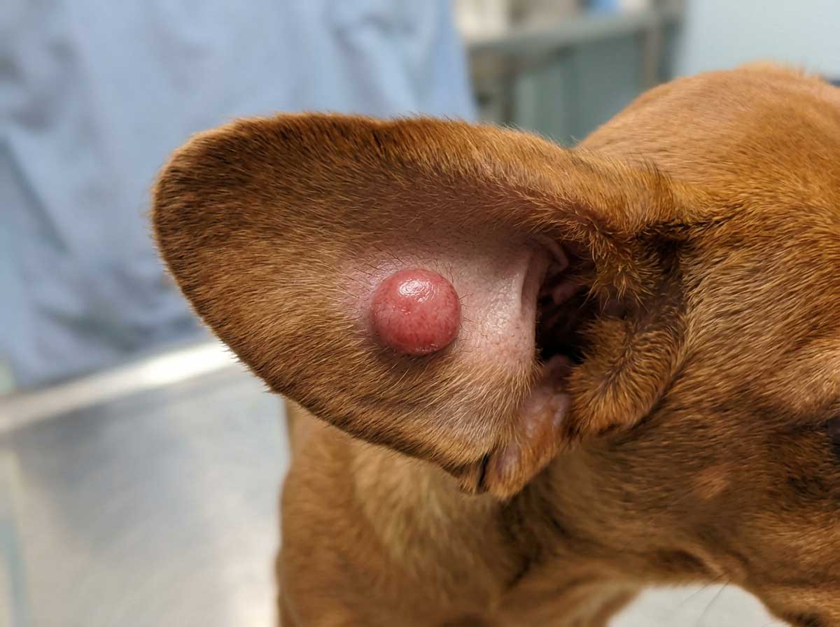
Histiocytoma (Button Tumor)
Histiocytomas are small, round, raised, red, hairless lumps that appear suddenly — often within just a few days. They look alarming (like a red button on the skin) but are benign tumors of immune cells. They're most common in dogs under 3 years old. The hallmark is a fast-appearing, dome-shaped, red lump usually on the head, ears, or legs. Despite their rapid appearance, histiocytomas typically resolve on their own within 2-3 months as the immune system destroys them. They're sometimes confused with mast cell tumors, so a vet check is recommended to confirm. No treatment is usually needed unless the lump doesn't resolve, ulcerates, or is in a location where it bothers the dog.
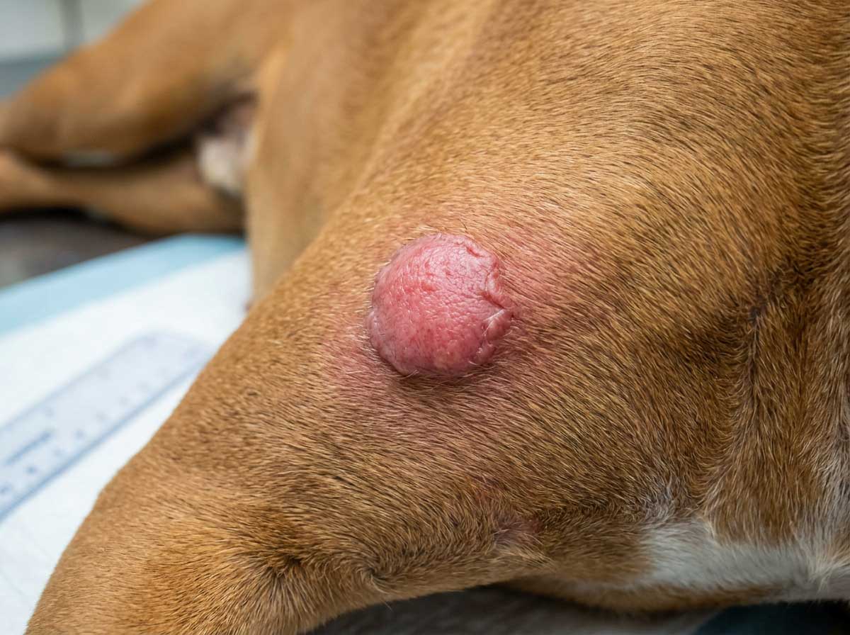
Mast Cell Tumor (Warning Signs)
Mast cell tumors (MCT) are the most common malignant skin tumors in dogs, accounting for about 20% of all skin tumors. The tricky part is they can look like almost anything — a small pink bump, a raised red lump, a wart-like growth, or even what appears to be a bug bite. Mast cell tumor dog symptoms include a lump that changes in size (swells and shrinks), redness or irritation around the lump, and sometimes surrounding skin hives. Breeds at higher risk include Boxers, Bulldogs, Boston Terriers, Golden Retrievers, and Labrador Retrievers. Dog histiocytoma vs mast cell tumor can be hard to distinguish visually. Any lump that changes size, grows rapidly, or doesn't resolve within 2-3 months needs a vet check and fine needle aspirate. Early detection and surgical removal dramatically improve prognosis.
Found a lump on your dog?
Upload a close-up photo of the lump or bump now. Get an AI-powered analysis to help you understand what it might be and whether to see a vet.
Check Dog Lump Now →Medical Disclaimer
PawCheck provides AI-generated preliminary health analysis for informational and educational purposes only. This service is not intended to replace professional veterinary advice, diagnosis, or treatment. The AI analysis has inherent limitations and may not always be accurate. Always seek the advice of a qualified, licensed veterinarian with any questions regarding your pet's health. Never disregard professional veterinary advice or delay in seeking it because of information provided by this tool. If your pet is experiencing a medical emergency, contact your veterinarian or emergency animal hospital immediately. By using this service, you acknowledge and agree to these terms.
Frequently Asked Questions
When should I worry about a lump on my dog?
+
Most lumps on dogs are benign (60-80%), but you should see a vet if: the lump is growing rapidly (doubling in size within weeks), it feels hard and immovable (fixed to underlying tissue), it's irregular in shape with uneven borders, it's ulcerated, bleeding, or oozing, your dog seems painful when you touch it, it appeared suddenly and is larger than a golf ball, or your dog has other symptoms like weight loss, lethargy, or appetite changes. Any new lump should ideally be checked by a vet — a simple fine needle aspirate (FNA) can often determine if it's benign or concerning.
How do I tell if my dog has a cyst or tumor?
+
Cysts and tumors can look similar from the outside, but there are clues. Cysts are typically round, smooth, well-defined, and may feel fluid-filled or slightly squishy. They often have a visible pore or opening and may leak thick, cheesy material if ruptured. Tumors tend to be more solid, may feel firmly attached to deeper tissue, and can be irregular in shape. However, the only reliable way to distinguish them is through veterinary testing — a fine needle aspirate (FNA) takes just minutes and examines the cells under a microscope. Don't rely on feel alone, as some cancers mimic benign cysts.
What do cancer lumps look like on dogs?
+
Cancerous lumps in dogs vary widely in appearance, which is why visual diagnosis alone is unreliable. Warning signs include: rapid growth over days or weeks, hard and firmly attached to underlying tissue (doesn't move freely), irregular or uneven borders, ulceration or bleeding on the surface, color changes (red, purple, black, or mottled), and size larger than 2cm. Mast cell tumors — the most common malignant skin tumor in dogs — can look like almost anything: a small pink bump, a raised red lump, or even a wart-like growth. This is why any new or changing lump should be tested by a vet rather than diagnosed by appearance.
Are dog tumors hard or squishy?
+
Both — and that's what makes them tricky. Lipomas (fatty tumors, benign) are typically soft, squishy, and move freely under the skin. Sebaceous cysts also feel soft or fluid-filled. However, some malignant tumors like mast cell tumors can also feel soft initially. Hard, immovable lumps are generally more concerning, but softness doesn't guarantee a lump is benign. The key warning signs are rapid growth, firmness that increases over time, attachment to deeper tissue (doesn't slide around), and changes in size or appearance. A vet can do a quick needle aspirate to check the cells regardless of how the lump feels.
How can you tell if a dog's lump is fatty or cancerous?
+
Fatty lumps (lipomas) are typically soft, round, move freely under the skin when you push them, grow slowly over months/years, and don't bother the dog. Cancerous lumps tend to be firmer, grow faster, may feel attached to deeper tissue, and can change in appearance. However, there's significant overlap — some cancers feel soft, and some lipomas feel firm. The only definitive answer is a fine needle aspirate (FNA), where your vet inserts a small needle, collects cells, and examines them under a microscope. It's quick, inexpensive ($50-$150), and usually doesn't require sedation. Never assume a lump is "just a fatty tumor" without testing.
How do you tell the difference between a skin tag and a tumor?
+
Skin tags are small, soft, flesh-colored or slightly darker, pedunculated (hang from a thin stalk), and stay the same size over time. They're extremely common in older dogs and completely benign. Tumors are typically broader-based (wide attachment to skin), may be firm or hard, can grow over time, and may change color or ulcerate. A tick can also be mistaken for a skin tag — look for tiny legs near the attachment point. If a "skin tag" starts growing, changes color, bleeds, or becomes firm, have your vet check it. Skin tags on the eyelid are common and usually harmless but should be monitored if they irritate the eye.
Are skin tags on dogs concerning?
+
Skin tags (acrochordons) on dogs are almost always benign and harmless. They're extremely common in older dogs, certain breeds (Cocker Spaniels, Poodles, Miniature Schnauzers), and overweight dogs. They don't need treatment unless they're in a location where they get caught or irritated (collar line, armpit, between toes), they bleed from being snagged, or they're growing or changing appearance. If a skin tag starts growing rapidly, changes color, becomes firm, or bleeds without being snagged, have your vet evaluate it — in rare cases, what looks like a skin tag can be an early-stage tumor.
How long will a dog live with a mast cell tumor?
+
Prognosis depends heavily on the grade and stage. Low-grade (Grade I) mast cell tumors have an excellent prognosis — with complete surgical removal, most dogs live a normal lifespan. Intermediate-grade (Grade II) tumors have variable outcomes; complete removal with clean margins often results in good long-term survival. High-grade (Grade III) tumors are aggressive and can spread to lymph nodes and organs; average survival with treatment is 6-12 months, though some dogs do better. Early detection and complete surgical removal are key. If you notice any lump on your dog that's growing, changing, or looks unusual, see your vet promptly — catching mast cell tumors early dramatically improves outcomes.
More AI Pet Health Checks

Eye Infection Checker
Detect dog eye infection, cat eye problems, conjunctivitis, corneal ulcers, and cataracts.

Skin Disease Checker
Detect dog skin infection, cat skin problems, dermatitis, hot spots, and ringworm.

Dog Ear Infection Checker
Detect dog ear infection, ear mites, yeast infections, and ear discharge with AI photo analysis.

Cat Ear Mites Checker
Detect cat ear mites, ear infections, yeast infections, and ear discharge with AI photo analysis.

Cat Vomit Checker
Why is my cat vomiting? Analyze white foam, yellow bile, blood, hairballs, and more with AI.

Dog Vomit Checker
Dog vomiting when to worry? Analyze white foam, yellow bile, blood, and undigested food with AI.

Dog Nose Checker
Dog nose dry or runny? Analyze cracked noses, nasal discharge, and color changes with AI.
Dog Dental Checker
Detect dog tooth infections, gum disease, tartar buildup, and dental problems with AI photo analysis.

Dog Poop Checker
Blood in dog stool? Analyze poop color, detect worms, mucus, and diarrhea with AI photo analysis.

Cat Dental Checker
Detect cat tooth infections, gum disease, stomatitis, tooth resorption, and dental problems with AI photo analysis.

Cat Poop Checker
Blood in cat stool? Analyze poop color, detect worms, mucus, and diarrhea with AI photo analysis.
Dog Wound Checker
Is your dog's wound infected? Check infection signs, healing stages, and bite wounds with AI photo analysis.

Cat Lump Checker
Found a lump on your cat? Identify skin tags, cysts, lipomas, warts, and bumps with AI photo analysis.

Cat Nose Checker
Cat nose dry or runny? Analyze crusty noses, nasal discharge, and sneezing symptoms with AI.
🐾
View All Tools
Explore all AI pet health check tools