Dog Hair Loss Checker — Bald Spots, Alopecia Pattern Photo AI
Dog losing hair in patches, on tail, around eyes, or with no itching? Upload a photo — AI identifies the PATTERN and LOCATION, ranks the most likely causes (ringworm, demodex, flea allergy, seasonal flank alopecia, or suspected endocrine disease), and flags honestly when blood work is needed to confirm.
Drop your pet's photo here
or
✅JPG, PNG, WEBP
📏Max 8MB
This tool provides AI-generated preliminary analysis only. Not a substitute for professional veterinary diagnosis.
📸 Photo Guide
Good photos

Close-up, fur parted

Shows border + healthy coat
Avoid

Too far away

Blurry / motion
Tips for best results
- ✓Part the surrounding fur so the AI can see where normal coat ENDS and bald area BEGINS
- ✓Include some healthy surrounding coat in the frame — comparison helps AI judge severity
- ✓For bilateral symmetric hair loss (flanks, body), take one photo of EACH side — helps confirm symmetry
- ✓For around-eye hair loss, photograph both eyes if affected — symmetric vs one-sided matters
- ✓Use natural daylight — it reveals skin color (reddened, darkened, or normal) most accurately
- ✓Note any scabs, scales, redness, or black "flea dirt" — capture them clearly if present
- ✓If the hair loss is in a BIG PATCH, take one close-up + one wider shot to show total area
- ✓Important honest note: AI reliably identifies visual patterns (location, shape, skin appearance). For suspected endocrine disease (Cushing's / hypothyroidism), the AI will recommend blood work — photo analysis alone cannot confirm these.
How It Works — AI Dog Hair Loss Pattern Checker

Step 1
Upload a Photo
Take a clear, well-lit photo of the bald or thinning area. Part the surrounding fur so the AI can see the border between normal and affected skin. Include a small area of healthy coat for comparison.

Step 2
AI Analyzes Pattern
Our AI reads the pattern (circular, symmetric, patchy), location (eyes, tail, flank, belly), and whether the skin is inflamed. It then ranks the most likely causes and flags when blood work is needed to confirm.

Step 3
Get Your Triage Report
Receive the pattern assessment, top 2-4 likely causes ranked by probability, urgency level (ER / vet this week / monitor), and honest flags when the photo alone cannot distinguish between causes that need blood work (like Cushing's vs hypothyroidism).
Dog Hair Loss Patterns — What Each Means
Dog hair loss has many causes. Our AI identifies the PATTERN (circular, symmetric, patchy, specific location) and narrows down the likely causes. Some patterns are visually clear (ringworm, flea allergy), others need blood work to confirm (endocrine disease). We're honest about both. Here's what the 8 most common patterns look like. Also try our dog skin checker or dog bug identifier or dog gum & tongue checker.
Circular Bald Patches with Scales (Ringworm Pattern)
Classic ringworm (dermatophytosis) creates round or oval hair loss patches, typically 0.5-5 cm across, with scaly, crusty edges. The center may be nearly bald, the border often reddish and flaky. Despite the name, ringworm is a FUNGAL infection, not a worm. It's common in puppies, kittens, immunocompromised animals, and dogs from kennels or shelters. IMPORTANT: ringworm is highly CONTAGIOUS to other pets and to humans — fungal spores persist in the environment for 18+ months. Diagnosis: woods lamp UV exam (about 50% of species fluoresce), fungal culture (definitive, takes 2-4 weeks), or PCR (fast). Treatment: topical antifungal shampoo (miconazole, ketoconazole) 2-3x per week AND oral antifungal (itraconazole, terbinafine) for 4-12 weeks, plus environment decontamination with dilute bleach. AI confidence for ringworm pattern: HIGH when classic circular with scaling is visible. Linked with /skin page for broader skin assessment.


Bald Patches with Scabs or Pustules (Bacterial Folliculitis / Pyoderma)
Bacterial folliculitis causes hair loss in patches, typically with small red pustules, scabs, or dried crusts visible on or around the bald areas. Often starts as a few spots and spreads. Common causes: secondary to allergies (scratching creates openings for bacteria), staphylococcus overgrowth, flea allergy, moist environment. The classic "dog losing hair in patches and scabs" query points to this. Signs include: "moth-eaten" appearance, circular expanding lesions (similar to ringworm but with more obvious pustules and redness), mild-to-moderate itching, sometimes a slight smell. Diagnosis: skin cytology (swab + microscope), bacterial culture if recurring. Treatment: 3-6 weeks of oral antibiotics (cephalexin, clindamycin, Simplicef) + medicated baths (chlorhexidine or benzoyl peroxide PET shampoo). Usually hair fully regrows after infection clears. Recurring folliculitis often points to underlying allergies or immune issues — workup recommended if it keeps coming back.
Hair Loss Around Eyes & Muzzle (Demodex / Allergies)
Hair loss concentrated around the eyes, muzzle, and paws most commonly suggests: (1) DEMODECTIC MANGE (demodex) — the #1 cause in puppies and young dogs under 18 months; small patches with slightly reddened skin, usually NOT itchy; skin scrape under microscope is diagnostic; localized demodex often self-resolves in healthy puppies, generalized demodex needs medication (Bravecto, NexGard). (2) ALLERGIES — food or environmental; typically bilateral, paired with itching, rubbing face on carpet/furniture; diagnosis requires elimination diet or allergy testing. (3) HYPOTHYROIDISM in older dogs — can produce a "mask-like" facial hair thinning. (4) Skin fold dermatitis in brachycephalic breeds (Pugs, Bulldogs, Shih Tzu) — bacterial/yeast in the skin folds around eyes and muzzle. AI pattern confidence: HIGH when classic demodex pattern with normal-to-slightly-red skin is present, especially in puppies. For adult-onset demodex, underlying immune suppression should be ruled out.


Tail Base / Rump Hair Loss (Flea Allergy Dermatitis)
Hair loss concentrated at the tail BASE (where the tail meets the back) extending across the rump is the CLASSIC pattern of flea allergy dermatitis. Even ONE flea bite can trigger massive hair loss in flea-allergic dogs — you may never see a flea. Look for flea dirt: tiny black specks that look like coffee grounds; place some on wet white paper towel — if they turn reddish-brown, it's digested blood = flea dirt. Pattern: hair loss at tail base + sometimes belly + inner thighs; intense itching; moderate-to-severe inflammation. Treatment: start prescription flea prevention IMMEDIATELY (NexGard, Credelio, Bravecto — all kill fleas within 4-8 hours); treat the house (adult fleas are only 5% of population — eggs and larvae everywhere); short-course steroid or Apoquel for severe itching; antibiotics if secondary skin infection. Hair typically regrows in 4-8 weeks after fleas are controlled. AI pattern confidence: VERY HIGH when tail-base concentrated hair loss + redness is present. Linked with /bug page to identify suspected fleas.
Bilateral Symmetric Flank Hair Loss, No Inflammation (Seasonal Flank Alopecia)
Seasonal Flank Alopecia (SFA) is a highly specific pattern: matching hair loss on BOTH flanks (sides of the body between ribs and hips), sharply demarcated, appears in late fall/winter, often regrows by spring. Critical distinguishing features: SYMMETRIC (both sides match), NO itching, NO redness, NO scaling, skin often DARKER (hyperpigmented) in the affected area but otherwise healthy. Predisposed breeds: BOXER (classic), English Bulldog, French Bulldog, Schnauzer (all sizes), Airedale, Scottish Terrier, Labrador, Bull Mastiff. Cause: poorly understood, thought to involve response to changes in daylight length affecting melatonin production. Treatment: often NONE needed — it's cosmetic. Melatonin supplementation (3-6 mg daily, vet-approved) can help some dogs. Light therapy (increasing daily light exposure) also tried. Hair often regrows naturally next spring/summer. BEFORE committing to this diagnosis, vet should rule out endocrine disease with bloodwork (thyroid, cortisol) since Cushing's/hypothyroidism can look similar. AI pattern confidence: HIGH when breed + symmetric bilateral flank pattern + seasonal onset + normal skin appearance present.


Symmetric Body-Wide Thinning, No Itch (Suspected Endocrine — NEEDS BLOOD WORK)
Symmetric, non-inflammatory hair loss spreading across the trunk, flanks, and belly — while the head and legs are often spared — points to ENDOCRINE DISEASE. Common causes and how they differ: (1) HYPOTHYROIDISM — symmetric hair loss; "rat tail" (bald tail); weight gain despite normal eating; lethargy; cold intolerance; dull thickened coat. Middle-aged dogs (4-10 yrs) of many breeds. Treated with daily thyroid hormone — inexpensive, very effective, hair usually regrows in 3-6 months. (2) CUSHING'S DISEASE (hyperadrenocorticism) — symmetric hair loss; thin fragile skin; pot-belly appearance; increased thirst/urination; panting; muscle weakness. Seniors 9-11 yrs, predisposed breeds: Poodle, Dachshund, Boxer. Treated with trilostane or mitotane — needs monitoring, responds well. (3) ADDISON'S DISEASE (hypoadrenocorticism) — less common cause of hair loss; more often presents with vomiting/weakness. (4) ALOPECIA X — cause unclear; typically affects plush-coated breeds (Pomeranian, Chow Chow, Keeshond, Siberian Husky, Malamute). IMPORTANT HONEST NOTE: AI PHOTO ANALYSIS CANNOT DISTINGUISH BETWEEN THESE ENDOCRINE DISEASES — they all look similar visually. If AI sees this pattern, it flags "suspected endocrine disease — blood work needed to confirm." A simple vet visit with blood tests (thyroid panel, ACTH stim for Cushing's) gives definitive diagnosis. Untreated endocrine disease causes long-term health problems beyond hair loss.
Older Dog Tail Hair Thinning ("Rat Tail" / Stud Tail)
Hair loss on the tail specifically — leaving the tail looking "bare," "rat-like," or with a greasy patch near the base — has specific causes in adult/senior dogs: (1) HYPOTHYROIDISM "rat tail" — classic presentation; entire tail hair thins; often combined with lethargy, weight gain, cold intolerance; highly treatable. (2) STUD TAIL (supracaudal gland hyperplasia) — greasy/waxy patch at the BASE (top) of the tail; caused by overactive sebaceous gland; most common in intact males, occasionally intact females; treatment is neuter + topical antiseborrheic shampoo. (3) AGE-RELATED thinning — mild gradual overall thinning in very senior dogs, symmetric, no skin change; usually doesn't require treatment. (4) TAIL TIP HAIR LOSS — from "happy tail" injury (tail hits walls), trauma, or vascular issues in small/old dogs. (5) BILATERAL tail + flank hair loss = endocrine disease (see previous section). AI pattern confidence: HIGH for stud tail (distinctive location + texture); MODERATE for "rat tail" appearance suggesting hypothyroidism — bloodwork recommended. Hair typically regrows with appropriate treatment.


Sudden All-Over Shedding (Stress / Post-Surgery / Nutritional)
Dramatic shedding over days to weeks — far more than seasonal shedding — can result from: (1) TELOGEN EFFLUVIUM (stress-induced) — major stressor (boarding, move, new pet, surgery, illness) pushes many hair follicles into rest phase simultaneously; massive shed 1-3 months AFTER the stressful event; regrows naturally over 2-4 months. (2) ANAGEN EFFLUVIUM — medication-induced (chemotherapy, some steroids) — hair in active growth phase dies; regrows after medication stops. (3) POST-SURGICAL hair loss at clip site — hair may not regrow for 3-12 months at surgical clip area; sometimes permanent in certain breeds. (4) NUTRITIONAL deficiency — rare with commercial diets, possible with unbalanced home-cooked diets; protein, zinc, and essential fatty acid deficiencies can cause diffuse hair loss. (5) SEASONAL BLOW — some breeds (Huskies, Malamutes, German Shepherds, Labradors) have twice-yearly "blows" where undercoat sheds dramatically; usually normal seasonal pattern but can look alarming; regrows within 2-3 weeks. (6) POST-PARTUM shedding in mother dogs — hormonal shift 2-3 months after whelping; regrows naturally. For sudden all-over shedding, the vet workup focuses on ruling out systemic illness (bloodwork) and confirming nutrition is adequate. Most cases self-resolve within 3-4 months.
Bald spots or unexplained shedding?
Upload a photo now — we identify the PATTERN and LOCATION, rank the most likely causes, and tell you honestly when blood work is needed to confirm. Not itchy? Not red? We can still help — that pattern is often the clearest diagnostic clue.
Check Dog Hair Loss Now →Medical Disclaimer
PawCheck provides AI-generated preliminary health analysis for informational and educational purposes only. This service is not intended to replace professional veterinary advice, diagnosis, or treatment. The AI analysis has inherent limitations and may not always be accurate. Always seek the advice of a qualified, licensed veterinarian with any questions regarding your pet's health. Never disregard professional veterinary advice or delay in seeking it because of information provided by this tool. If your pet is experiencing a medical emergency, contact your veterinarian or emergency animal hospital immediately. By using this service, you acknowledge and agree to these terms.
Frequently Asked Questions
Why is my dog losing hair in patches but not itchy?
+
Non-itchy hair loss in patches points AWAY from the most common inflammatory causes (flea allergy, bacterial infection, sarcoptic mange) and TOWARDS: (1) Ringworm — circular patches with scaling, often not itchy especially in early stages; highly contagious to humans and other pets. (2) Demodectic mange — especially in puppies and around eyes/muzzle; causes patchy hair loss with mild or no itching. (3) Endocrine disease — Cushing's, hypothyroidism, Addison's; usually symmetric bilateral pattern with normal-looking skin underneath. (4) Seasonal flank alopecia — bilateral flank patches, no skin changes, common in Boxer/Bulldog/Schnauzer. (5) Bacterial folliculitis in early stage — patches may have scabs but minimal itching initially. (6) Post-injection alopecia — at the site of a previous vaccine or injection. Non-itchy hair loss still needs a vet — the lack of itching actually narrows possibilities but doesn't mean it's benign. Blood work is usually the next step (rules out endocrine), plus a skin scrape for mites and fungal culture for ringworm.
Why is my dog losing hair all of a sudden?
+
Sudden hair loss ("it wasn't like this last week") can be triggered by: (1) Stress — post-boarding, new home, surgery, or loss of a companion can cause "telogen effluvium" — hair cycling into rest phase and shedding en masse 1-3 months after the stressful event. (2) Allergic reaction — food, new medication, new collar/harness material, environmental (pollen). (3) Flea infestation that reached critical mass. (4) Ringworm exposure (daycare, dog park, new puppy in home) — incubation ~2 weeks then sudden onset. (5) Post-surgical clipping alopecia — hair at the site where they shaved for surgery may not regrow for 3-6 months or longer. (6) Anagen effluvium from certain medications (chemotherapy, some steroids). If the hair loss is dramatic and happening fast, see a vet within a week. Take a photo of the affected area for comparison over time.
What does Cushing's hair loss look like in dogs?
+
Cushing's disease (hyperadrenocorticism) causes a distinctive pattern: SYMMETRIC bilateral hair loss that usually starts on the TRUNK and FLANKS, spreads to the sides and belly, but typically SPARES the head and legs. The skin underneath often appears THIN, DARK (hyperpigmented), and sometimes shows calcinosis cutis (calcium deposits — gritty white patches). Unlike inflammatory hair loss, the skin is usually NOT red and the dog is NOT itchy. Additional Cushing's signs: pot-belly appearance (abdominal fat redistribution), increased thirst/urination, panting excessively, muscle weakness, weight gain. Diagnosis requires blood tests (ACTH stimulation or low-dose dexamethasone suppression test) plus sometimes abdominal ultrasound. Breeds predisposed: Poodle, Dachshund, Boxer, Boston Terrier, Beagle, Yorkshire Terrier. Seniors most commonly affected. AI photo check CAN identify the suspicious pattern, but CANNOT confirm Cushing's vs other endocrine diseases — bloodwork is essential.
Can alopecia in dogs be cured?
+
Depends entirely on the cause. Most cases are manageable or fully curable with proper diagnosis: (1) FULLY CURABLE: ringworm (antifungals + environment cleaning), flea allergy (flea control), localized demodex in healthy puppies, bacterial folliculitis (antibiotics). (2) MANAGEABLE: Cushing's (daily medication like trilostane — hair regrows as disease is controlled), hypothyroidism (thyroid hormone replacement — hair often fully regrows in 3-6 months). (3) SELF-RESOLVING: seasonal flank alopecia (often regrows next season). (4) HARDER TO TREAT: generalized demodex in immunocompromised dogs, Alopecia X (unclear cause), post-clipping alopecia (may take 6-12+ months). (5) USUALLY NOT REVERSIBLE: pattern baldness in certain breeds (Chihuahua pinnal alopecia, Dachshund body pattern), scarring alopecia from previous severe disease. Getting the right diagnosis is key — same-looking hair loss can have opposite treatments. Don't try "cure-alls" before knowing the cause.
Will my dog's bald spots grow back?
+
In most cases YES, if the underlying cause is treated. Regrowth timelines: (1) Flea allergy — 4-8 weeks after fleas are controlled. (2) Ringworm — 6-12 weeks after antifungal treatment starts. (3) Bacterial infection — 4-8 weeks after antibiotics finish. (4) Hypothyroidism — 3-6 months after starting thyroid hormone replacement. (5) Cushing's — 4-8 months after medication starts controlling cortisol. (6) Seasonal flank alopecia — often regrows in next winter/seasonal cycle. (7) Post-surgical clipping alopecia — variable, 3-12 months; some dogs never fully regrow the hair. (8) Stress-induced (telogen effluvium) — 2-4 months after the stressor resolves. Cases where hair may NOT regrow: long-standing untreated disease causing follicular scarring, breed-specific pattern baldness (genetic), extensive damage from self-trauma. The sooner you identify and treat the cause, the more complete the regrowth.
How do I treat my dog's hair loss at home?
+
Home treatment depends on knowing the cause — which is why AI photo check + vet evaluation come FIRST. That said, here are safe supportive measures once you know the cause: (1) FLEA-RELATED: start prescription flea prevention (NexGard, Credelio, Bravecto), wash bedding on hot, treat the house. (2) RINGWORM: bathe with ketoconazole/miconazole shampoo (pet-formulated, not human); disinfect surfaces with dilute bleach; keep affected pet separated. (3) ALLERGIES: eliminate suspect food/material; add omega-3 fish oil (1000mg per 20 lbs body weight daily) — reduces skin inflammation over 6-8 weeks. (4) DRY SKIN: omega-3, humidifier in winter, avoid over-bathing. (5) GENERAL SUPPORT: oatmeal baths soothe irritation; do NOT apply human products (Neosporin is potentially toxic if licked, benzoyl peroxide only in pet formulations). WHAT NOT TO DO: don't use human shampoo, human hair regrowth products (Rogaine/minoxidil is TOXIC to dogs — can be fatal), essential oils, or hydrogen peroxide on hair loss sites. Endocrine diseases have NO home remedy — medication is required.
What does it mean when a dog loses hair around the eyes?
+
Hair loss around the eyes is a specific pattern that narrows causes significantly: (1) DEMODECTIC MANGE (demodex) — the #1 cause in puppies and young dogs; often starts around eyes or muzzle as localized demodex; patches of hair loss with slightly reddened skin; usually not itchy; diagnosed by deep skin scraping. (2) ALLERGIES — environmental or food allergies can cause hair loss around eyes due to rubbing; often paired with itching and face rubbing on carpet/furniture. (3) HYPOTHYROIDISM — can cause hair loss in a "rat-tail" pattern or around the face/eyes in middle-aged to senior dogs. (4) CONJUNCTIVITIS / tear staining — chronic eye discharge can lead to hair loss around eyes as moisture damages fur. (5) BACTERIAL INFECTION — secondary to allergies or skin fold dermatitis (common in Pugs, Bulldogs, Shih Tzu). (6) RINGWORM — less common but possible; usually circular and scaly. (7) SELF-TRAUMA from painful eye condition (corneal ulcer, glaucoma). A vet visit is standard for eye-area hair loss — rule out mange with a scrape, rule out eye pain with a fluorescein stain.
Why is my dog missing lots of fur on her tail?
+
Tail fur loss has specific patterns pointing to specific causes: (1) TAIL BASE / RUMP area — classic flea allergy dermatitis; even if you don't see fleas, one bite can cause massive fur loss here in allergic dogs. Check for flea dirt (tiny black specks). (2) TAIL TIP (the very end) — "happy tail" injury from repeatedly hitting walls, trauma, or circulation issues. (3) WHOLE TAIL with "rat tail" appearance — classic hypothyroidism pattern in middle-aged/senior dogs; skin underneath may be darker; combined with weight gain and lethargy. (4) TOP OF TAIL with greasy/waxy texture — "stud tail" (supracaudal gland hyperplasia); common in intact males, sometimes intact females; sebaceous gland overactivity. (5) UNDER tail + around anus — anal gland issues, licking from discomfort. (6) SYMMETRIC bilateral tail hair loss — could be endocrine disease. (7) SUDDEN fur loss at tail base WITH skin change — could be bacterial folliculitis or pyoderma. Most tail hair loss is treatable once the cause is identified — check for fleas FIRST since that's the most common.
Will coconut oil help my dog's hair grow back?
+
Short answer: probably not significantly, but it's unlikely to hurt if used cautiously. Coconut oil contains medium-chain fatty acids that MAY have mild antimicrobial properties and provide some moisturizing — but there's little scientific evidence it speeds hair regrowth in dogs. What actually helps: (1) TREATING THE UNDERLYING CAUSE (most important — no topical will work if thyroid is low or mange is active). (2) Omega-3 supplementation (fish oil) — actual evidence for improving coat and skin over 6-8 weeks. (3) Proper nutrition with adequate protein. (4) Regular flea prevention. Risks of coconut oil: dogs often lick it off (promotes ongoing licking of the hair loss area — counterproductive); can cause digestive upset if a lot is ingested; a few dogs are allergic. Better topical options if your vet approves: pet-specific medicated shampoos, pet-safe moisturizers recommended for the specific cause. Don't use as a substitute for actual treatment — if hair loss persists 2+ weeks on home remedies, see a vet.
Do dogs lose hair on their tail as they age?
+
Some age-related tail hair thinning IS normal in senior dogs, but dramatic hair loss is NOT just "aging" — there's usually a cause worth investigating. Normal aging changes: slight overall coat thinning, some grayscale/white hair increase, slightly drier skin, modest decrease in overall fur density. These should be symmetric and gradual over months to years. NOT normal (even in seniors): patches of hair loss, "rat tail" appearance (common in hypothyroidism — very treatable!), bald spots, thinning that happens over weeks instead of months. Senior dogs are actually MORE prone to specific hair loss causes: Cushing's disease (peak age 9-11), hypothyroidism (peak age 4-10), skin tumors, and stud tail in intact males. If your senior dog has noticeable tail hair loss, it's worth a vet visit and bloodwork — hypothyroidism in particular is extremely common, highly treatable, and often causes dramatic improvement in coat, energy, and weight within months of starting thyroid medication.
What does seasonal flank alopecia look like in dogs?
+
Seasonal flank alopecia (SFA, also called "cyclical flank alopecia") is a distinctive, mostly cosmetic condition with a very specific appearance: (1) BILATERAL SYMMETRIC hair loss — matching patches on BOTH flanks (sides of the body between ribs and hips). (2) Sharply demarcated borders — the bald areas have clear edges, not gradual fading. (3) Underlying skin is often DARKER (hyperpigmented blue-gray) than surrounding skin but usually NOT red, not swollen, not itchy. (4) Appears MOST commonly in late fall/winter (November-April in northern hemisphere); regrows in spring/summer. (5) May recur year after year in affected dogs. Breeds strongly predisposed: Boxer (classic presentation), English Bulldog, French Bulldog, Schnauzer, Airedale, Scottish Terrier, Labrador. Treatment options: often NONE needed (cosmetic only); melatonin supplementation (3-6 mg daily, vet-approved) helps some dogs regrow hair faster; lightbox therapy (increasing light exposure). Confirmed by appearance + breed + seasonal pattern; ruling out endocrine disease with bloodwork is standard. Most affected dogs live completely normal lives — it's a cosmetic issue in otherwise healthy dogs.
What breeds get seasonal flank alopecia?
+
Seasonal flank alopecia most strongly affects: BOXER (the classic breed — probably the most frequently diagnosed), ENGLISH BULLDOG, FRENCH BULLDOG, SCHNAUZER (Miniature, Standard, Giant), AIREDALE TERRIER, SCOTTISH TERRIER, BULL MASTIFF, LABRADOR RETRIEVER (less common but reported), GOLDEN RETRIEVER (occasionally), STAFFORDSHIRE BULL TERRIER, and AMERICAN BULLDOG. Less commonly: Great Dane, American Bulldog, Doberman, Rhodesian Ridgeback. Mixed breeds with these parents can also develop it. The underlying cause is thought to be a response to changes in daylight length (photoperiod), affecting melatonin production. If your dog is one of these breeds and develops bilateral symmetric flank hair loss in autumn/winter without itching or other symptoms, SFA is the most likely diagnosis — though your vet will still rule out endocrine disease with bloodwork before committing to this diagnosis.
Is alopecia in dogs dangerous or contagious?
+
Depends on the cause: DANGEROUS — hair loss ITSELF isn't dangerous, but some CAUSES are: (1) Cushing's disease — untreated, can lead to diabetes, blood clots, infections, decreased lifespan. (2) Hypothyroidism — untreated, causes weight gain, heart issues, reduced quality of life. (3) Addison's disease — can cause acute crisis (collapse, shock); treatable. (4) Underlying cancer causing paraneoplastic alopecia — the cancer itself is dangerous. NOT DANGEROUS: (1) Seasonal flank alopecia — purely cosmetic. (2) Post-clipping alopecia — cosmetic. (3) Pattern baldness — cosmetic. CONTAGIOUS causes: (1) RINGWORM — highly contagious to other pets AND humans; fungal spores persist in environment for 18+ months; requires treatment + environmental decontamination. (2) SARCOPTIC MANGE — contagious to dogs and can briefly affect humans. (3) FLEAS — the fleas themselves are contagious to other pets. (4) BACTERIAL INFECTIONS — occasionally contagious between pets. NOT CONTAGIOUS: allergies, endocrine disease, demodectic mange (lives normally on skin), seasonal flank alopecia, stress-induced hair loss. Photo assessment + vet visit helps sort this out quickly.
My dog is itching and losing hair but has no fleas — what could it be?
+
Itchy hair loss without visible fleas points to: (1) Flea allergy with hidden fleas — ONE flea bite can cause weeks of itching in allergic dogs; you may never see the flea. Do 2-week flea treatment trial before ruling out. (2) Environmental allergies (atopic dermatitis) — pollen, dust mites, mold; often seasonal worsening; affects face, paws, belly most. (3) Food allergies — often causes year-round itching including face, ears, paws, belly; takes 8-12 week elimination diet to diagnose. (4) Sarcoptic mange (scabies) — intense itching, often ear edges, elbows, and belly; highly contagious; needs skin scrape diagnosis. (5) Contact dermatitis — new detergent, lawn chemical, shampoo, collar material. (6) Bacterial skin infection — secondary to allergies; pustules + hair loss + itching. (7) Yeast infection (Malassezia) — greasy, smelly, itchy skin + hair loss; often in skin folds. (8) Ear mites (more common in cats but dogs affected) — intense head shaking + hair loss around ears. Next steps: vet exam + skin scraping + fungal culture + possibly allergy testing. In the meantime, start monthly flea prevention even if you haven't seen fleas — the most common "no fleas" scenario is actually "hidden flea allergy."
What does the first stage of alopecia look like in dogs?
+
Early-stage alopecia varies by cause, but common early patterns include: (1) THINNING BEFORE BALDNESS — fur looks sparser or less full in an area before visible skin shows through. Owners often describe it as "his coat looks weird but I can't pinpoint why." (2) DULLNESS and BREAKAGE — hair shafts weaken, fur looks dry/frizzy/broken before actually falling out (common in early endocrine disease). (3) HAIR PULLS OUT EASILY — running your hand through the coat and getting more hair on your palm than usual. (4) SMALL BALD AREAS — initially coin-sized, progress over weeks to months. (5) CHANGE AT SPECIFIC LOCATIONS first — around eyes (demodex, allergies), tail base (flea allergy), flanks (endocrine/SFA), paws (allergy/yeast). (6) COLOR CHANGE — some dogs develop darker skin color in the areas before hair loss becomes visible. (7) ITCHINESS BEFORE HAIR LOSS — inflammatory causes (allergies, infections) often itch first, then hair falls out from scratching. (8) SHEDDING PATTERN CHANGE — abnormal amounts or abnormal timing (not seasonal shedding). Take a photo when you first notice anything unusual — comparing against this baseline photo makes progression (or regrowth after treatment) much easier to judge.
More AI Pet Health Checks

Eye Infection Checker
Detect dog eye infection, cat eye problems, conjunctivitis, corneal ulcers, and cataracts.

Skin Disease Checker
Detect dog skin infection, cat skin problems, dermatitis, hot spots, and ringworm.

Dog Ear Infection Checker
Detect dog ear infection, ear mites, yeast infections, and ear discharge with AI photo analysis.

Cat Ear Mites Checker
Detect cat ear mites, ear infections, yeast infections, and ear discharge with AI photo analysis.

Cat Vomit Checker
Why is my cat vomiting? Analyze white foam, yellow bile, blood, hairballs, and more with AI.

Dog Vomit Checker
Dog vomiting when to worry? Analyze white foam, yellow bile, blood, and undigested food with AI.

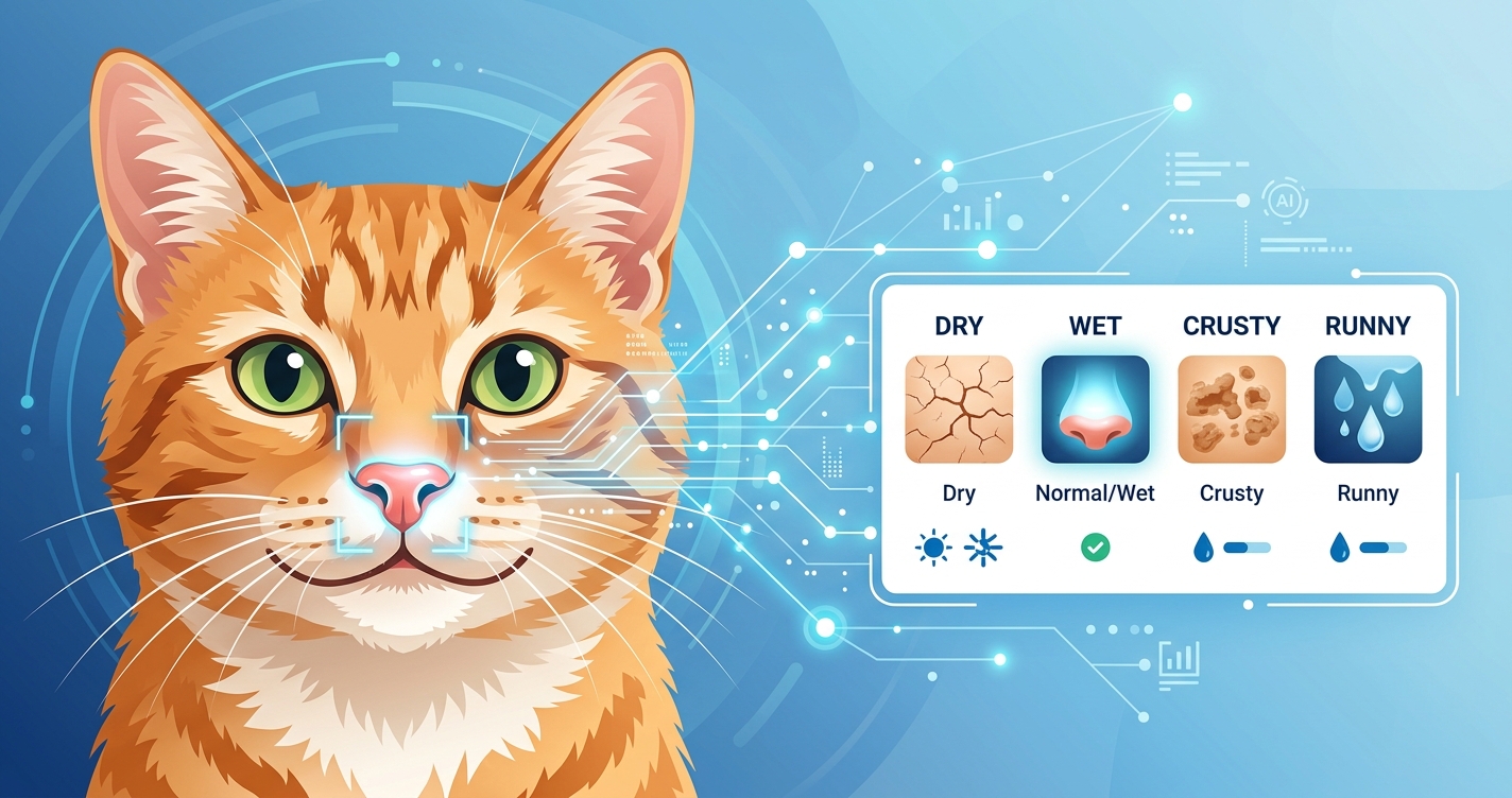
Dog Nose Checker
Dog nose dry or runny? Analyze cracked noses, nasal discharge, and color changes with AI.
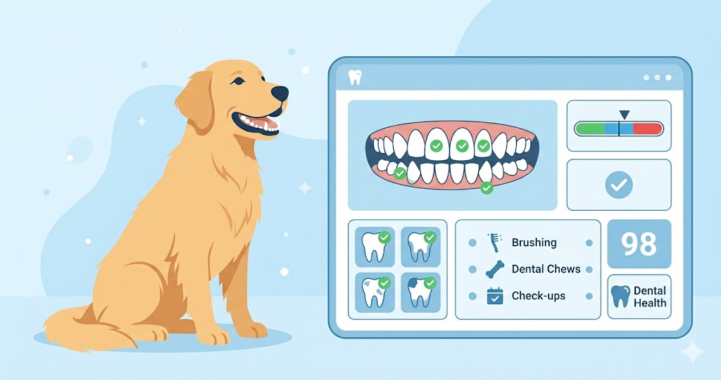
Dog Dental Checker
Detect dog tooth infections, gum disease, tartar buildup, and dental problems with AI photo analysis.

Dog Poop Checker
Blood in dog stool? Analyze poop color, detect worms, mucus, and diarrhea with AI photo analysis.
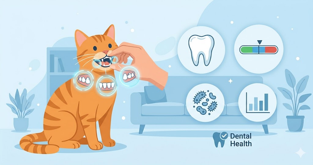
Cat Dental Checker
Detect cat tooth infections, gum disease, stomatitis, tooth resorption, and dental problems with AI photo analysis.

Dog Lump Checker
Found a lump on your dog? Identify skin tags, warts, cysts, lipomas, and bumps with AI photo analysis.

Cat Poop Checker
Blood in cat stool? Analyze poop color, detect worms, mucus, and diarrhea with AI photo analysis.
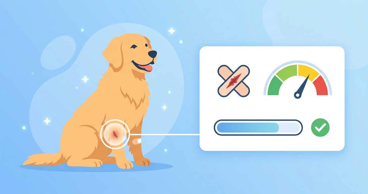
Dog Wound Checker
Is your dog's wound infected? Check infection signs, healing stages, and bite wounds with AI photo analysis.

Cat Lump Checker
Found a lump on your cat? Identify skin tags, cysts, lipomas, warts, and bumps with AI photo analysis.
Cat Nose Checker
Cat nose dry or runny? Analyze crusty noses, nasal discharge, and sneezing symptoms with AI.

Dog Paw Checker
Dog paw injured, swollen, or infected? Check cuts, yeast infections, peeling pads, and redness between toes with AI.

Dog Acne Checker
Pimples on your dog's chin or muzzle? Tell canine acne from mange with AI — includes puppy acne, severity stage, and treatment advice.

Dog Urine Checker
Blood in your dog's urine? Dark or orange pee? Check for UTI, dehydration, liver issues, or emergency signs with AI photo analysis.

Cat Paw Checker
Cat paw swollen, puffy, or injured? Check pillow foot, infections, abscesses, ingrown nails, and pad problems with AI.

Cat Acne Checker
Black specks on your cat's chin? Tell feline acne apart from flea dirt or mites with AI — includes severity stage and treatment advice.

Cat Urine Checker
Blood in your cat's pee? Dark or cloudy urine? Check color, clarity, and visible blood with AI — triage UTI, crystals, or liver issues.

Dog Gum & Tongue Checker
Pale, blue, yellow gums or black spots on tongue in your dog? Triage anemia, bloat, jaundice, toxin exposure, or benign lentigo with AI photo analysis.

Cat Gum & Tongue Checker
Pale, blue, yellow, or red gums/tongue in your cat? Triage FeLV, feline asthma, stomatitis, jaundice, or toxin exposure with AI photo analysis.

Cat Hair Loss & Overgrooming Checker
Cat licking fur off, losing hair on belly, or scruffy coat? AI identifies miliary dermatitis, stud tail, ringworm, or flags paraneoplastic cancer warning in senior cats.

Dog Broken Nail Checker
Dog broken nail bleeding, hanging, exposed quick, or infected? AI assesses severity and gives step-by-step home treatment or clear vet-visit guidance.

Dog Eye Discharge Checker
Green, yellow, clear, or brown eye discharge? AI identifies the color and ranks causes — bacterial infection, allergies, dry eye, porphyrin tear stains, or foreign body.

Cat Eye Infection & Discharge Checker
Green, yellow, brown, watery, or black crust eye discharge? AI identifies feline herpesvirus, chlamydia, URI, bacterial infection, or blocked tear duct — with urgency triage.
🐾
View All Tools
Explore all AI pet health check tools